
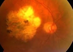
Long term anti-VEGF treatments (injections for wet macular degeneration) raises concern about retinal atrophy!
If you are receiving injections for macular degeneration you should reconsider! Over 18% of patients reviewed in this national study developed retinal atrophy which is a more severe form of macular degeneration. Atrophy is a severe condition of the death of the retinal cells.
I am not surprised to read the results of this study because injections go against homeopathic laws of healing. In order to properly treat macular degneration you must look for the underlying cause of the disease! According to Herrings Law, if you just suppress the symptoms of wet macular the disease will be pushed deeper into the body and a more serious condition will develop, i.e., retinal atrophy.


18.3% of CATT Patients (Comparison of Age-related macular degeneration Treatment Trials) developed geographic atrophy.
Comparison of Age-related macular degeneration Treatment Trials (CATT)
■ One of the great unresolved questions to come out of the recent, large-scale CATT trial comparing the safety and efficacy of ranibizumab (Lucentis, Genentech, South San Francisco, CA) with bevacizumab (Genentech) was whether repeated dosing of these anti-VEGF therapies over a period of time could trigger geographic atrophy (GA) in patients who had shown no signs of GA at baseline.
A number of respected retina specialists, including Philip Rosenfeld, MD, PhD, of Bascom Palmer Eye Institute in Miami, FL, have been leaning toward the position that repeated anti-VEGF over time can trigger GA. However, Dr. Rosenfeld and others wanted to see additional studies that might provide confirmation of this hypothesis.
Now, a team of researchers led by Juan E. Grunwald, MD, of the University of Pennsylvania, has published a study of 1,024 CATT patients whose color fundus photos or fluorescein angiograms showed no visible signs of GA at enrollment. After two years, the researchers found that GA had developed in 187 patients, or 18.3% of the cohort being studied.
While the researchers noted several possible factors that could have contributed to the development of GA, including lower visual acuity at baseline, retinal angiomatous proliferation, and foveal intraretinal fluid, they also implicated monthly dosing and the use of ranibizumab in GA development. They concluded that “anti-VEGF therapy may have a role in the development of GA.”
“This study confirms the suspicions previously raised,” says Dr. Rosenfeld. “This new analysis is all very consistent with prior reports. The information I want to see is whether the appearance and growth of GA in the HARBOR study was influenced by monthly dosing or the higher Lucentis dose. These are very important data.”
REFERENCE
Grunwald JE, Daniel E, Huang J, et al. Risk of geographic atrophy in the Comparison of Age-related Macular Degeneration Treatments Trials (CATT). Ophthalmology. 2013 Sep 30. [Epub ahead of print]
Retina. 2014 Jul;34(7):1308-15.
Exacerbation of choroidal and retinal pigment epithelial atrophy after anti-vascular endothelial growth factor treatment in neovascular age-related macular degeneration.
Young M1, Chui L, Fallah N, Or C, Merkur AB, Kirker AW, Albiani DA, Forooghian F.
PURPOSE:
To study the progression of retinal pigment epithelium (RPE) and choroidal atrophy in patients with neovascular age-related macular degeneration (AMD) and to assess for a possible association with the number and type of anti-vascular endothelial growth factor treatments.
METHODS:
Patients with neovascular AMD and a minimum of 1-year follow-up were reviewed. Fellow eyes with nonneovascular AMD were used as control eyes. Retinal pigment epithelial atrophy area and choroidal thickness were determined using spectral-domain optical coherence tomography. Multivariable regression models were used for statistical analyses.
RESULTS:
A total of 415 eyes were included in the study, with a mean follow-up of 2.2 years. Eyes with neovascular AMD had greater progression of RPE atrophy and choroidal atrophy compared with those with nonneovascular AMD (P < 0.001). Progression of RPE atrophy and choroidal atrophy was independently associated with the total number of injections of bevacizumab and ranibizumab (all P values ≤ 0.001). In the subgroup of 84 eyes with neovascular AMD and without RPE atrophy at baseline, only bevacizumab was associated with the progression of RPE atrophy (P = 0.003). This study likely lacked statistical power to detect an association with ranibizumab in this subgroup.
CONCLUSION:
Retinal pigment epithelial atrophy and choroidal atrophy in neovascular AMD seem to be exacerbated by anti-vascular endothelial growth factor treatment. Possible differences between bevacizumab and ranibizumab require further investigation.
http://www.ncbi.nlm.nih.gov/pubmed/24451923